

An atlas of life: mapping the human body cell by cell
The Human Cell Atlas is one of the most ambitious projects in modern biology. By building a map of every cell type in the human body, researchers across the world are transforming how we understand health and disease.

Licence: Attribution CC BY
13-minute read
13-minute read
Elapsed time:00:00Total time:00:00
“Contemplate the heartbeat: this phenomenon that many of us might consider the epitome of the everyday – the heart will beat more than two billion times over an average person’s life – is, in fact, a miraculously complex feat of cell biology. The heart is a model of cellular cooperation, citizenship, and belonging.”
“The way I think about a tissue is that it’s an ecosystem of cells that are constantly talking to each other,” says Sarah Teichmann, co-founder of the Human Cell Atlas (HCA). “It’s that community that delivers the function.”
This principle is true at every scale: “Different tissues in an organ work together,” says Sarah. “So, if we take the heart, there are about 30 different tissues – the wall, the valves, the vessels, the conduction system – that work together to deliver the heartbeat. Those interactions are absolutely crucial.”
Sarah’s view of biology could just as well describe the Human Cell Atlas initiative itself. It is a community – on a global scale – of diverse experts and specialists working towards a common goal. The heart is just one of 18 biological maps that make-up the Human Cell Atlas.
Since 2016, more than 4,000 scientists from more than 100 countries have contributed. Biologists, clinicians, technologists, data scientists, and many others – all working together to achieve one thing: a comprehensive profile of the identity and location of every cell type in the human body.
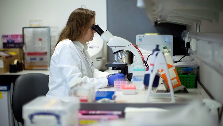
Sarah Teichmann co-founded the Human Cell Atlas, a global effort to map the identity and location of every cell type in the human body.
Watch: Mapping every cell type in the human body
Pressing play on the video above will set a third-party cookie. Please read our cookie policy for more information.
Muzlifah Haniffa and Sarah Teichmann introduce the Human Cell Atlas, a global effort to map the cells of the human body. They explain how single-cell and spatial genomics, together with organoids, are deepening our understanding of human biology, health and disease.
Muzlifah Haniffa
When you plant a seed in the soil, it's that interaction between the seed and the soil that allows that seed to then grow into a little plant. Two things come together, interconnect and it forms something different. And that interdependency is what our cells need to form these structures that makes the human body. How do these cells come together? How do they build a tissue? And how do they build a human?
Sarah Teichmann
So there are approximately 37 trillion cells in the human body. The $64,000 question is how many cell types are there? Answering that question is what the Human Cell Atlas project is all about. The way I think about a tissue is that it's an ecosystem of cells that are constantly talking to each other, and it's that community that delivers the function of the tissue. And the beauty of the Human Cell Atlas is that we can understand those cellular conversations that are taking place within a tissue, down to the molecular level. The typical Cell Atlas project has multiple stages. And of course, the first stage is actually gathering the tissue samples.
Researcher 1
I'm going to take my forceps. I'm just going to place it into the metal. And then I'm just going to fill the whole thing up with wax, so it embeds nicely. And then I'm just going to put it on this side to freeze. And then it'll freeze into a nice wax block. And then we continue with the sectioning. So this is a five micron thick section. So then we can put it onto a slide. And then we can look at the cells and the ecosystem we see within this small section.
Muzlifah Haniffa
My clinical specialty is in dermatology. That led me to dissect the cells that make up the skin. And over time, I realised that genomics held an enormous potential. What you can see here is the top layer of the skin. But now, with the technology and what the Human Cell Atlas is doing is the ability to view these tissues not in the shades of pink and purple. Now, with the spatial technology, we've now got all of the individual cell types that are in the skin being shown here. Every cell in the tissue, we have an identity for and we know the gene expression of these cells. And you will see that the hair follicle is not just made up of one cell type. There are many different cell types. This is where the stem cells are and the base of the hair follicle from which the shaft of the hair grows out of. And so it's absolutely incredible that we can now look at the resolution of individual cells and their gene expression.
Sarah Teichmann
This is a massive project. And in terms of the scale of the data, we've sequenced over 100 million cells. And we're going to scale up to a billion. And achieving that has been a collaboration between over 4,000 scientists around the world from over 100 countries. So this is a global community that's working together to build a map of the human body.
Muzlifah Haniffa
If we can understand how cells are formed and how they come together, the opportunities are enormous. You can then work out how to build those cells. Can you make a tissue that you can use for transplant? Can you make hair follicles that can induce hair formation? The beauty of organoids is you can actually watch this in real time. An organoid is a collection of cells. And you can make an organoid from an inducible pluripotent stem cell, an embryonic stem cell, or you can take patient tissue and derive an organoid from that.
Researcher 2
Organoids can be made across a variety of different tissue types. So I'm just focusing the microscope so I can see the different details of the skin organoid. We can now see skin developing in the dish in real time. Every hair follicle in our body develops before we are born. So with conditions that affect hair loss, maybe understanding how these hair follicles develop we can then better inform therapeutic strategies to improve outcomes for patients.
Muzlifah Haniffa
When I saw the first skin organoid, I was flabbergasted. I could not believe how well they resembled human skin in our body. There are multiple skin diseases. Let's take, for example, hair loss. We can profile those tissues, but what you get are serial snapshots. You can't watch it as a video reel. So with an organoid now, we can actually take the patient's tissue and then look to prevent or revert disease. And you can test that in a dish. And mechanistic understanding is what we really need in order to find new therapeutic strategies and ways to prevent disease.
Sarah Teichmann
There are other organoid systems that mimic the heart, parts of the brain, endometrial organoids that mimic the monthly menstrual cycle. Organoids are incredibly useful for validating discoveries from the Human Cell Atlas and can be used to predict drug effects, side effects, etc. The Human Cell Atlas data has very immediate tangible biomedical relevance, and so the impact has been very fast and very deep and cuts right across academia, industry, health care, public health. In order to make the data available, the funding from sources like the Wellcome Trust has been absolutely crucial, allowing everyone across the world to benefit from this data, from this project. It's been a tremendous pleasure and an honour to lead this community, and has really been a very special journey and a very special part of my scientific career.
A decade of insights
In the 10 years since the HCA was launched, this effort has yielded important, clinically relevant insights.
Researchers have so far analysed 2.3 million of the heart’s cells. Discoveries have included identifying the rare pacemaker cells that set its rhythm. “They’re a clock that beats on a second-by-second basis,” says Sarah. The researchers found that GLP-1 drugs – used to treat obesity and diabetes – could act directly on these cells, helping explain a slight increase in heart rate seen in clinical trials.
During the COVID-19 pandemic, Atlas researchers rapidly examined which cells expressed the receptor used by SARS-CoV-2 to enter the body. This highlighted potentially vulnerable cells in the nose, mouth and eye.
Atlas data is also showing where and when genes linked to developmental disorders are active. “We can now map to where the genes that cause developmental disorders are expressed, exactly where in space and exactly when in time,” says Muzlifah Haniffa, Deputy Director of the Wellcome Sanger Institute and co-chair of the Human Cell Atlas. This could help clinicians identify which organs may be affected and inform investigations and longer-term care.
“The impact has been very fast and very deep,” says Sarah. “[It] cuts right across academia, industry, healthcare and public health.”
When the project began, just over a decade ago, achieving these results seemed impossibly ambitious.
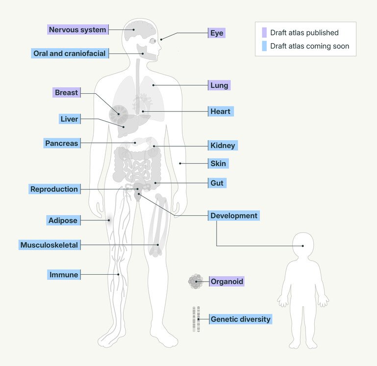
The Human Cell Atlas’s Biological Networks combine data from multiple studies to build integrated reference maps of human cells. The colours show progress towards publishing these draft atlases.
Selected insights emerging from the Human Cell Atlas
Drug effects on the heartbeat: Mapping specialised pacemaker cells helped show that GLP-1 may increase heart rate by acting directly on the sinus node.
Treatment opportunities for the lung: Researchers found shared cell states across pulmonary fibrosis, cancer and COVID-19, pointing towards possible common therapeutic targets.
Origins of inflammatory bowel disease: Mapping the gut from early development to adulthood revealed processes that may contribute to Crohn’s disease and identified potential targets for treatment.
Clues for better healing: Mapping prenatal skin has shown that immune cells contribute to scarless healing, offering clues for regenerative medicine and treatments to prevent scarring.
Understanding joint disease: A developmental skeletal atlas linked distinct early bone and cartilage cells to later risk of hip and knee arthritis.
How reproductive organs form: Mapping the developing reproductive system revealed key genes and signals involved in organ formation, offering clues to the origins of congenital conditions.
Where the Human Cell Atlas began
The Human Cell Atlas was co-founded in 2016 by Sarah and Aviv Regev. Working separately on opposite sides of the Atlantic, their research had convinced them that mapping every cell in the human body was both important and increasingly possible.
Aviv had been at the Broad Institute in the US, developing techniques to analyse gene sequences in healthy and diseased cells. When Sarah became Head of the Cellular Genetics Programme at the Wellcome Sanger Institute, the two joined forces.
The official launch took place in October 2016 at Wellcome in London, where Sarah and Aviv brought together nearly 100 scientists. Over two days, the group began shaping the scientific roadmap and building an international consortium founded on openness, inclusion and the responsible sharing of data.
In an interview last year Sarah recalled that, at first, the prospect of "sequencing all the cells in the human body" seemed "slightly ridiculous". The Wellcome Sanger Institute's tradition of landmark endeavours – most notably the Human Genome Project – helped make the idea seem achievable.
“The Human Cell Atlas is the child or the progeny of the Human Genome Project,” says Muzlifah. “Sanger is where we first sequenced the genetic blueprint of what makes us human,” she adds. “That legacy is incredible.”
The team set out to build on this precedent by revealing how that blueprint is expressed in individual cells. They also aimed to capture a broader picture of human diversity. “At the very start, we wanted the Atlas initiative to be globally representative, so that we could actually profile cells and tissues of many individuals from all around the world,” says Muzlifah.
The work is organised through Biological Networks focused on particular tissues, organs and systems. There are now 18, and together they are assembling a first draft of the Atlas.
Through illuminating the function of specific cell types, the hope is to generate a deeper, broader understanding of how tissues, organs and living systems function in health and disease.
By mapping all the cells in the human body – and understanding how they change over time – the Human Cell Atlas is tackling one of biology’s most ambitious grand challenges. The project has built a vibrant global community, working openly and in partnership. It represents discovery science at its best.
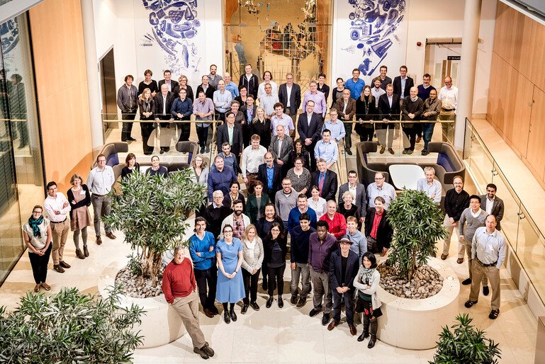
The Human Cell Atlas launched at Wellcome in London on 13-14 October 2016, bringing together experts from around the world to shape the project.
A spatial understanding of biology
How will the Atlas achieve this ambition? And how does understanding the molecular profiles of cells – and their locations – represent such a step forward in biology? The answer begins with how cells use their genes.
Almost every cell in the body contains essentially the same genome, but different genes are switched on in different cells. This means that, despite sharing the same genetic information, cells develop specialised identities and functions.
This development is influenced by many factors, including signals from the cell’s surrounding environment. These signals can affect which genes are switched on or off, guiding the cell towards a particular identity and function. “It’s the juxtaposition of cells coming together that creates something new,” says Muzlifah. She compares it to planting a seed. “It’s the interaction between a seed and the soil… that allows the seed to then grow into a little plant.”
Until recently, however, examining cells and their surroundings at this level of detail was extremely difficult. Before single-cell technologies, researchers generally took a tissue sample and broke open all its cells at once, analysing the resulting mixture of genetic material.
The result was an average signal across thousands or millions of cells, concealing rare cell types and important differences between them. The process also lost information about where individual cells had been located within the tissue.
Researchers can now study individual cells in molecular detail, examining gene activity and, increasingly, the proteins they contain. Spatial technologies capture this molecular information while preserving the cells' locations within a tissue. Combined, such data allows researchers to build detailed maps of how tissues are organised and, ultimately, how they react in different circumstances.
“The beauty of the Human Cell Atlas is that we can understand those cellular conversations that are taking place within a tissue down to the molecular level,” says Sarah. “We can not only get a much deeper understanding of how we work, but we can also have much better drug development, understanding of disease mechanisms, and then have huge impacts on human health.”
But the first draft of the Atlas will be only a starting point.

The Wellcome Sanger Institute helped establish the Human Cell Atlas, building on its role in the Human Genome Project.
Human Cell Atlas 2.0
The next stage will aim to map cells from more people around the world, across different cell states and stages of life. It will also add richer spatial information, moving towards full three-dimensional reconstructions of tissues and organs.
AI will likely be central to the Human Cell Atlas’ future, helping to integrate cellular data with genetics, imaging, and clinical information. Biology has entered an era of prediction, enabled by new computational and machine learning approaches that can analyse vast, complex datasets and use them to project forward in time. The revolution that is happening at the intersection of AI and biology is being powered by large-scale foundational data like the Human Cell Atlas.
“My hope for the future of the Human Cell Atlas Project is that we’ll achieve a full map of the human body at cellular resolution,” says Sarah. “And also that we will model that cellular data in a way that helps us for public health, global health, making medicines safer and more effective, developing new diagnostics and make a healthier future for the world and people everywhere.”
Organoids: building tissues in a dish
One of the HCA’s Biological Networks focuses on organoids: laboratory-grown models that resemble human tissues. By comparing organoids with cells from the human body, researchers can assess how accurately they reflect real tissues and identify ways to improve them.
The HCA provides a reference for understanding what organoids should look and behave like as they develop. This can help researchers refine the conditions in which organoids are grown and assess how closely they resemble the corresponding human tissue.
“It’s like having a recipe book that will guide you to how you should feed the organoid and how you should assess and benchmark the organoids against it with human tissue,” says Muzlifah. When she first saw a skin organoid, she was “flabbergasted”.
“I could not believe how well they resembled human skin in our body. It even had hairs, which was a bit creepy.”
By understanding how cells organise themselves into tissues, researchers hope to build increasingly realistic models for studying disease, testing drugs and advancing regenerative medicine.
Timeline of the Human Cell Atlas
Aviv and Sarah join forces, convening nearly 100 scientists in London, where the international Human Cell Atlas consortium takes shape.
The consortium publish the project’s first plan, form international networks and begin developing shared methods and pilot tissue maps.
Cell data helps researchers identify where the COVID-19 virus may enter the body. Early organ maps, including the lung, reveal previously unknown cell types. By December 2021 the project had more than 2,000 members from 77 countries.
The project continues to grow, expanding its datasets and bringing together thousands of scientists from more than 100 countries, with growing attention to ethics, equity and representation.
More than 45 papers mark major progress towards a first draft, including new maps of organs, blood vessels and human development at different life stages.
Human Cell Atlas and United Nations Educational, Scientific and Cultural Organization (UNESCO) announce a new collaboration to advance open science.
By April the project has more than 4,000 members from 103 countries. In October it marks its 10th anniversary.
Researchers continue combining the maps, adding data from more populations and developing better tools for exploring the atlas, including AI-based approaches.
Tags
Related content

